Quick access

Resources

Clinical insights, research, and resources for ultrasound innovation.

Publications

Explore clinical third-party publications and filter by the topics most relevant to you.

Explore more

Downloads

White papers, DICOM files, and training videos designed for clinical teams. Download what you need.

Explore more

News

Stay up to date with our latest press releases, product launches, and industry insights.

Explore more

FAQ

Find quick answers about
PIUR Infinity's functionality,
training and support.

Explore more

What the Scientific Literature Shows

Clinical publications

8 min read

Can Diabetologists Use Artificial Intelligence to Screen for Thyroid Nodules?
Hossam A Ghazi , Hanan A. Elsherbiny , Basma Z. Elmorsy , Amira S. Nowara
Anatomy-informed 3D Reconstruction of Tracked Ultrasound Sweeps

8 min read

Anatomy-informed 3D Reconstruction of Tracked Ultrasound Sweeps
Alexander Furtner, Zoe Reinke & Thomas Wendler

8 min read

Training the diagnostic artificial intelligence in thyroid sonography: how well is deep learning truly learning?
Oana Lozan, Petra B. Musholt, Ann-Kathrin Lederer, Nabila Bouzakri, Hauke Lang & Thomas J. Musholt
Validation studies

8 min read

An Ex Vivo Evaluation Of Tomographic 3-D Ultrasound, B-Mode Ultrasound, CT And MR Imaging To Measure Artery Diameter, Length And Wall Volume
By Steven Rogers, Joao Carreira, Ruth Thompson, Ana Morais, Christopher Miller, Wolfgang Wein, Jonathan Ghosh, Charles Mccollum
Validation studies

8 min read

Reliability And Accuracy Of Tomographic 3-D Ultrasound For Grading Vessel Stenosis: A Phantom Study
By Adel Alzahrani, Y Sultan A. Alotaibi, Mohammed Aslam, Salahaden R. Sultan
Validation studies

8 min read

Validation of a scanning technique with minimal compression for measuring muscle volume with freehand 3D ultrasound
By J. Huet, A.-S. Boureau, A. Sarcher, C. Cornu, A. Nordez

Access Exclusive Insights

DICOM Files

Explore real patient scan data from our PIUR tUS Infinity - Thyroid Application.

Access expert resources

Register to download practical reimbursement guides, whitepapers and more.

For our customers

Training videos

Step-by-step video tutorials to help you get started, optimize workflows, and make the most of PIUR Infinity in daily practice.

Watch on YouTube

User manuals

Comprehensive documentation covering installation, configuration, and clinical use of PIUR tUS Infinity.


User Manual | English
User Manual | US English
Gebrauchsanweisung | Deutsch
Manual de usuario | Español
Manuel d’utilisation | Français
Manuale d’uso | Italiano
User Manual | English
Gebrauchsanweisung | Deutsch
Manual de usuario | Español
المستخد م دليل | عربي
Manuel d’utilisation | Français
Manuale d’uso | Italiano
Podręcznik użytkownika | Polski
Manual do usuário | Português
lanıcı Kılavuzu | Türkçe

Marketing materials

Ready-to-use materials to support communication with patients, partners, and referring physicians.

PIUR tUS Infinity | English
PIUR tUS Infinity | Deutsch
Keyboard Shortcuts | English
Keyboard Shortcuts | Deutsch

Statements

DICOM Conformance Statement

Updates

Latest news and announcements

PIUR IMAGING Lands Former Sonosite CEO and Ultrasound Visionary Kevin Goodwin as Strategic Advisor
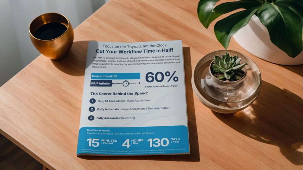
PIUR IMAGING Launches AI-Powered Software Reducing Thyroid Ultrasound Workflow Time by up to 60%
PIUR IMAGING Secures €5.6 Million Growth Capital to Scale Tomographic 3D Ultrasound in the USA and Europe
PIUR IMAGING Receives FDA Clearance for PIUR tUS inside, Bringing Advanced 3D Ultrasound to U.S. Healthcare
PIUR IMAGING Receives FDA Clearance for Revolutionary 3D Ultrasound Technology, PIUR tUS Infinity, Now Available in the U.S.

Frequently Asked Questions About PIUR

Everything about PIUR in one place — from how tomographic 3D ultrasound works to clinical applications, workflow, integration, pricing, and reimbursement. Grouped by topic for quick navigation.

Understanding PIUR Infinity

What is PIUR tUS Infinity and how does it work?

PIUR tUS Infinity is a tomographic ultrasound software solution that adds 3D imaging capabilities to conventional ultrasound systems. It converts acquired ultrasound image data into digital 3D volumes for review and analysis. PIUR Infinity also uses AI-based algorithms and machine learning to automate segmentation, measurements, annotations, and reporting workflows, helping standardize and streamline imaging processes.

Tomographic ultrasound (tUS) imaging is a method of acquiring and reconstructing ultrasound data into a navigable 3D volume rather than individual 2D images. By capturing a more complete anatomical dataset of the scanned region, tUS enables clinicians to review structures in 3D, perform measurements and annotations, and digitally store scans for longitudinal assessment.

PIUR Infinity transforms acquired ultrasound image data into navigable 3D volumes using reconstruction technology. Instead of relying on individual 2D images, clinicians can review complete volumetric datasets, visualize anatomy across multiple planes, and perform measurements, annotations, and longitudinal assessments within a digital 3D environment.

PIUR Infinity addresses challenges associated with conventional ultrasound workflows, including operator dependency, workflow inefficiencies, and limited access to complete imaging datasets. By enabling standardized 3D acquisition, digital storage, and AI-assisted workflow support, PIUR Infinity helps streamline image acquisition, support longitudinal follow-up, and reduce variability across users.

Unlike conventional ultrasound systems, PIUR Infinity is not an ultrasound machine but a software solution that works with compatible ultrasound platforms. PIUR Infinity adds navigable 3D imaging, AI-assisted workflow support, and digital volume storage to conventional ultrasound, enabling standardized acquisition, automated measurements, and separation of image acquisition from clinical review.

Most AI-based ultrasound solutions focus primarily on image analysis or nodule classification. PIUR Infinity, in contrast, supports the broader imaging workflow by combining 3D imaging with AI-assisted automation of measurements, annotations, and structured reporting. Rather than analyzing selected images alone, PIUR Infinity standardizes and streamlines the entire workflow from image acquisition and assessment through to documentation and follow-up.

Clinical Applications

What clinical applications can PIUR Infinity support?

PIUR Infinity supports multiple clinical applications through application-specific software licenses. Current available application licenses include thyroid imaging, as well as carotid plaque and vascular imaging workflows. Each application is optimized for its respective clinical use case and available feature set.

PIUR Infinity was initially developed with a strong focus on thyroid imaging and includes features such as automated lobe and nodule measurements, AI-assisted annotations, ACR TI-RADS calculator, structured reporting, and longitudinal follow-up support. While thyroid remains its primary and most advanced application, PIUR Infinity also supports additional clinical applications through application-specific software configurations.

PIUR Infinity supports additional clinical applications through application-specific software licenses. Current applications include thyroid imaging as well as selected vascular workflows such as carotid plaque assessment. Features and workflow capabilities may vary depending on the selected application.

Workflow & User Experience

How does PIUR Infinity fit into an existing ultrasound workflow?

PIUR Infinity integrates into existing ultrasound workflows as a software solution. Image acquisition is performed on compatible ultrasound systems, while analysis is carried out on a dedicated workstation where 3D volume reconstruction, AI-assisted measurements, annotations, and structured reporting are completed for clinical review and documentation.

Yes. Image acquisition with PIUR Infinity can be performed by trained nurses or technicians on compatible ultrasound systems, depending on local clinical protocols. Physicians do not need to be present during acquisition and can instead review the reconstructed 3D volumes, perform analysis, and complete reporting on a separate workstation.

Image acquisition with PIUR Infinity is typically completed in 30 seconds for thyroid imaging, with a single sweep per lobe capturing the full organ volume. Unlike conventional ultrasound workflows, analysis tasks such as measurements, nodule segmentation, annotations, and reporting are performed separately on a workstation after acquisition. This combination of workflow separation and automation of post-processing tasks enables a faster and more streamlined overall examination, showing up to 60% reduction in total exam time.

PIUR Infinity reduces physician workload by separating image acquisition from analysis, allowing scans to be performed by trained staff while physicians review the examination findings on a separate workstation at a later time, similar to a radiology review process. The system also automates routine tasks such as nodule segmentation, measurements, annotations, and structured reporting, reducing manual effort and improving overall workflow efficiency.

Technical Compatibility & Integration

Which ultrasound systems and manufacturers are compatible with PIUR Infinity?

PIUR Infinity is compatible with most major ultrasound systems from leading manufacturers, including GE, Philips, Siemens, Canon, and others. The system is designed to integrate with existing ultrasound infrastructure without requiring replacement of your machine.

Yes, PIUR Infinity is available both as a standalone add-on solution for most ultrasound systems and as an integrated application on select GE HealthCare LOGIQ systems under the PIUR inside configuration. The integrated version runs directly on the ultrasound platform, enabling streamlined 3D acquisition and analysis within the same system environment.

PIUR Infinity supports conventional B-mode ultrasound imaging data, which is reconstructed into navigable 3D volumes using tomographic ultrasound (tUS) technology.

Yes. PIUR Infinity uses a lightweight add-on hardware setup to enable 3D image acquisition without replacing existing ultrasound systems. This includes a sensor attached to the ultrasound transducer via a bracket, and a video box that captures live ultrasound images from the system's video output and transmits them to a workstation for reconstruction and analysis.

Yes. PIUR Infinity can export structured 3D datasets and reports to PACS systems, enabling integration with existing clinical storage and review infrastructure. This allows clinicians to access and review historical examinations within established PACS workflows without disrupting current processes.

Yes. PIUR Infinity supports export of DICOM-compatible data. This ensures that examination data can be stored, accessed, and reviewed within standard hospital workflows.

Purchasing, Pricing & Implementation

How much does PIUR Infinity cost?

PIUR Infinity is offered through a subscription-based model with a fee per examination. Pricing is typically tiered based on clinical volume, allowing clinics to scale costs according to patient throughput and usage levels. Specific pricing depends on configuration, application licenses, and expected examination volume.

You can request a demo through the 'Request Online Demo' page on our website. The demo typically includes an overview of PIUR Infinity, a walkthrough of the workflow from image acquisition to 3D analysis and reporting, and a discussion of clinical applications, integration options, and implementation setup.

The PIUR Infinity system includes software for 3D tomographic ultrasound (tUS) imaging and analysis, clinical application-specific licenses, and a lightweight hardware setup consisting of a sensor with a transducer bracket and a video box.

Yes. PIUR IMAGING provides installation support to ensure proper setup and integration of PIUR Infinity into existing ultrasound workflows. This includes assistance with hardware installation (sensor, bracket, and video box), software configuration, and system integration with the ultrasound device and workstation environment. Remote and on-site support are available depending on the implementation.

Yes. PIUR IMAGING provides onboarding and training to support clinical adoption of PIUR Infinity. This includes guidance on image acquisition, software use on the workstation, and interpretation of 3D datasets within structured clinical workflows.

Yes. PIUR IMAGING provides technical support to assist with system setup, integration, and ongoing use of PIUR Infinity. Support is available for both software and hardware components, including troubleshooting, workflow questions, and system maintenance, ensuring continuous clinical operation.

Reimbursement & Economics

Can clinics receive additional reimbursement for examinations using PIUR Infinity?

Yes. When PIUR Infinity is used in thyroid imaging workflows, clinics may be able to report CPT 76377 (3D rendering with image post-processing) in addition to CPT 76536 for standard thyroid ultrasound, depending on clinical documentation and payer requirements. This can result in additional reimbursement of approximately $80–$200 per examination.

PIUR Infinity supports the use of Category I CPT codes for thyroid ultrasound workflows, including CPT 76536 for standard thyroid imaging and CPT 76377 for 3D rendering with image post-processing performed on a dedicated workstation. These codes may be used together depending on clinical documentation and payer requirements.

Clinics typically evaluate ROI based on three main factors. First, time savings from automated measurements, segmentation, and reporting workflows. Second, increased reimbursement potential through eligible Category I CPT codes. Third, improved workflow efficiency through standardized acquisition, which can reduce repeat examinations and variability in image acquisition quality. Together, these factors can contribute to higher clinical throughput and improved operational efficiency.

Still need help?

Reach out to our team directly.

Contact us

FREE Whitepaper

Don’t settle for inconsistent thyroid measurements.

Discover how upgrading to 3D ultrasound imaging can reduce variability in volume measurements and elevate your clinical precision.​
Download your free copy today and learn why leading clinicians are choosing 3D imaging for more reliable, accurate thyroid assessments.