Last updated: 16 October 2026
Clinically Reviewed by Charlsie McDonald, RDMS (AB)(BR)


The scan is the part everyone sees. The reporting is where the time goes. This article follows ultrasound reporting from the probe going down to the patient hearing the result.
Written for sonographers, and for the department heads deciding whether the workflow is worth changing.
You know the sequence. The friction hides in the joins.
The patient is positioned and acquisition begins. Measurements are taken live, on the machine, with the probe still on the patient. Which images get saved is not a free choice: every facility works to a protocol, set by itself or its accrediting body, defining the minimum image set for each study. For thyroid, that typically means annotated sagittal and transverse views of each lobe and the isthmus. Those go to PACS. Measurements either flow into the worksheet through a structured reporting link or get typed in by hand. You add a technical comment, and the study leaves your hands.
Somewhere else, later, a reading physician opens it, works from the images you saved, and dictates or enters the report. Someone reviews, someone corrects, someone signs. It lands in the RIS or EHR. Then it keeps moving: the signed report goes to the ordering physician, who is the one who speaks to the patient. Sometimes a portal shows the report before that conversation happens.
The ultrasound machine, PACS, the RIS or structured reporting tool, speech recognition, the hospital information system. Five systems, several people, a dozen handoffs between the scan and the signed sonography report. Any one of them can add a day.
Some of it is the scan. Most of it is not.
The scan itself, when the exam is difficult. Straightforward studies move quickly. Difficult anatomy, a large habitus or an uncooperative patient can stretch an examination considerably: in our experience, a demanding thyroid study can take 30 to 45 minutes.
Retyping. Where the ultrasound system and the reporting tool are not linked, numbers are read off one screen and typed into another. Fast per exam, and easy to stop noticing.
Correcting the report afterwards. Dictation-based reporting is not a clean channel. A 2015 systematic review of speech recognition in radiology departments found that, in the mostly older studies it covered, the proportion of reports containing at least one error ranged from 4.8% to 89% for speech recognition, and from 2.1% to 22% for transcription.
Source: Hammana I et al., Speech Recognition in the Radiology Department: A Systematic Review, Health Information Management Journal 44(2):4-10, 2015.
Rescanning, when something was not captured. The protocol protects against most of this: if the required views are defined, they get taken. What it cannot catch is a finding nobody saw. On a fast-moving list, a small lesion can go unnoticed, and an unnoticed lesion is not imaged, not measured, and not in the record to go back to.
Waiting. The study sits in a worklist until a reading physician opens it, and the signed report still has to reach the ordering physician before the patient hears anything. None of that is in the sonographer’s hands.
Underneath most of that sits one structural fact: in conventional freehand 2D ultrasound, what was not captured does not exist.
The protocol guarantees a minimum. It does not guarantee a record. The saved images are a defined set plus whatever the sonographer noticed in the moment and thought to document, assembled before anyone knew which question would end up mattering. You watched the nodule come in and out of view and formed an impression from something dynamic. Nobody downstream sees any of that. They see the protocol images and your comment.
That is why the reading physician has to ask. And why a scan that was never saved can mean calling the patient back, where a stored volume can often be revisited instead.
The consequence is measurable. In a phantom study co-authored by PIUR IMAGING and comparing multiplanar 2D and 3D methods for thyroid volumetry, conventional B-mode 2D ultrasound showed strong observer dependence, with operator experience having the most pronounced effect, while all three tracked 3D methods, including PIUR’s, reduced inter- and intraobserver variability. A separate study in 28 healthy volunteers, using the PIUR tUS system with the authors’ own research segmentation pipeline, found that tracked 3D ultrasound significantly reduced interobserver variability compared with conventional 2D.
Sources: Roth R et al., Tomographic 3D Ultrasound for Thyroid Volumetry: A Comparison Between Multiplanar 2D and 3D Imaging Methods, Diagnostics 16(15):2329, 2026. Krönke M et al., Tracked 3D ultrasound and deep neural network-based thyroid segmentation reduce interobserver variability in thyroid volumetry, PLOS ONE 17(7):e0268550, 2022.
Store the sweep as a 3D volume rather than a set of frames, and the sweep itself becomes the record.
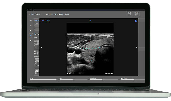
PIUR tUS Infinity does this with a sensor mounted on a standard ultrasound probe: you scan on the system you already have, and the software reconstructs the tracked freehand sweep into a tomographic volume.
Three things follow:
Measurement becomes retrospective. Volumes and dimensions can be derived from the stored sweep after the patient has left, including ones nobody thought to take at the time.
Acquisition becomes a sweep. For thyroid, documenting the anatomy means one sweep of about 30 seconds per lobe rather than capturing and annotating image after image.
The reading physician can navigate the volume rather than working from a fixed set of frames, which is what separating acquisition from analysis means in practice.
Source: PIUR IMAGING, Thyroid Imaging.
Clinicians describe the effect in terms of what the time gets spent on instead. Dr. Edouard Ghanassia, of the Thyroid Diagnosis and Interventional Center in Sète, France, puts it this way:
With PIUR, I have more time for medical reasoning, anatomical particularities, and diagnosis, instead of spending it on standard measurements.
Source: PIUR IMAGING testimonials.
Independent research on freehand 3D ultrasound in general, not specific to PIUR tUS Infinity, points the same way. In a prospective clinical trial in 14 patients, using postoperative specimens as the reference standard, median volume difference from the true value was 1.7 mL for 3D ultrasound versus 3.6 mL for 2D (p = 0.02). Inter-operator reliability for 3D ultrasound was 0.986, where 1.0 would be perfect agreement between operators.
Source: Rask KB et al., Thyroid Volume Measurement With AI-Assisted Freehand 3D Ultrasound Compared to 2D Ultrasound: A Clinical Trial, Head & Neck 48(1):64-70, 2026.
For thyroid work, computer-assisted volume measurement and an ACR TI-RADS calculator sit on top of the volume as decision support: the physician confirms the classification, and the numbers entering the report come from the stored data rather than from a keyboard.
The output has to land where the report actually gets written.
PIUR tUS Infinity works with ultrasound systems from GE, Philips, Siemens, Canon and other manufacturers that provide a digital video output. Patient reports and imaging data are automatically generated, PowerScribe compatible, and sent directly to PACS, so they arrive in the dictation environment most departments already run. The physician still reviews and signs the report.
PIUR tUS inside is available on GE HealthCare’s LOGIQ™ Open Platform Technology as a hosted application on LOGIQ™ E10, LOGIQ™ E10s, and LOGIQ Fortis™ systems, enabling 3D acquisition and analysis directly on the ultrasound platform. Contact your local GE HealthCare sales representative for availability.
Source: PIUR IMAGING, Platform and Thyroid Imaging.
Two questions are worth putting to your own IT team: what the DICOM output contains in your configuration, and whether your specific PACS is validated or simply compatible. The answers differ by site, and they decide how much of the workflow above you actually get.
PACS compatibility. What format does your PACS accept, and do volumes arrive as reviewable objects or flattened captures?
Where measurements land. Automatically in the worksheet, or is someone still typing?
Protocol fit. Your accreditation protocol defines a required image set. Confirm what the volume-based workflow submits against it.
Who signs what. If a measurement is taken retrospectively, who owns it in the signed report? A governance question, not a technical one.
Template carry-over. Templates and macros usually survive, but the fields they expect must match what the new workflow produces.
Training time. A sweep is a different motor skill from targeted 2D. Ask how many scans before sweeps are reliably usable.
A pilot with clear limits. One scanner, one indication, a fixed number of exams, and a measure everyone agreed on before you started.
If you would like to see how a single sweep turns into a report on the ultrasound system you already use, our team is happy to walk you through it with your own protocol in mind.
Short answers to the questions sonographers and department heads ask most often before moving from a 2D image set to a stored 3D volume.
No. The examination still depends on a sonographer covering the anatomy and on the judgment that spots what the protocol never asked for. What changes is the manual side of documentation: instead of capturing and annotating image after image, you sweep once and work from the volume.
That depends on your department’s governance and on the regulatory status of the software in your region. Settle it before a pilot, not after.
No. PIUR tUS Infinity combines software with a small sensor that attaches to your existing probe. It works with systems from GE, Philips, Siemens, Canon and other manufacturers that provide a digital video output.
In published studies, yes. In a clinical trial in 14 patients, freehand 3D ultrasound measured thyroid volume more accurately than the 2D ellipsoid method, with an inter-operator reliability of 0.986 (Rask et al., Head & Neck, 2026; research on freehand 3D ultrasound in general). A volunteer study using the PIUR tUS system found significantly lower interobserver variability than conventional 2D (Krönke et al., PLOS ONE, 2022).
Reach out to our team directly.
Contact usThis article is provided for general informational purposes for healthcare professionals. It does not constitute medical advice and does not replace clinical judgment, local protocols or the instructions for use. Results from independent research cited here do not refer to PIUR IMAGING products unless stated otherwise, and outcomes in individual settings may differ. Product availability, intended purpose, and regulatory status vary by jurisdiction. Please contact PIUR IMAGING for information on your region. PowerScribe is a trademark of Nuance Communications, Inc., a Microsoft company. All other product and company names are trademarks of their respective owners.
FREE Whitepaper
Don’t settle for inconsistent thyroid measurements.
Discover how upgrading to 3D ultrasound imaging can reduce variability in volume measurements and elevate your clinical precision.
Download your free copy today and learn why leading clinicians are choosing 3D imaging for more reliable, accurate thyroid assessments.
